This article was first published by doctors Miloud Chakit, PhD, Rachid Ayoub Zahir, MD, Abdelhalem Mesfioui, PhD on Elsevier journal www.elsevier.com/locate/radcr
Miloud Chakit, PhDa,∗, Rachid Ayoub Zahir, MDb, Abdelhalem Mesfioui, PhDa
a Biology and Health laboratory, Faculty of Sciences, Ibn Tofail University, Kenitra Morocco
Head of Urology Department, El Idrissi Hospital, Kenitra, Morocco
a r t i c l e i n f o
Article history:
Received 4 January 2024
Revised 12 March 2024
Accepted 18 March 2024
Keywords: Nephrectomy Pyonephrosis Urolithiasis Diabetes Case report
a b s t r a c t
Pyelonephritis is one of the main systemic bacterial infections encountered in emergency departments. We present a case of diabetes woman aged 30 years referred to our urology department of El-Idrissi Hospital, Kenitra (Morocco) for recurrent episodes of urinary tract infection, multiple urolithiasis, chills, unilateral lower back pain, chills and severe hydroureteronephrosis. Abdominal CT showed a non-functioning obstructed kidney with pyelic and ureteral stones. Nephroureterectomy was performed by extraperitoneal nephrec- tomy for avoiding any more extended nephrectomy incision or second iliac incision, this technic ensures nephroureterectomy with minimal risk of affecting the distal ureter, that sometimes follows nephrectomy.
Diabetes and urolithiasis coexistence in a patient may cause severe pyonephrosis leading to nephroureteroctomy.
© 2024 The Authors. Published by Elsevier Inc. on behalf of University of Washington. This is an open access article under the CC BY-NC-ND license (http://creativecommons.org/licenses/by-nc-nd/4.0/)
Introduction
Pyonephrosis is a severe infection related to a retention of pus in the dilated urinary tract with destruction of the renal parenchyma. The causes are generally renal damage, per- sistent urinary obstruction, urolithiasis and urinary system infection [1]. The incidence of pyonephrosis is higher in older female patients and affects mostly the left kidney [2]. Several causes are related to its aetiology like urinary tract infection,
urolithiasis, urinary obstruction, lipid metabolism abnormal- ities, lymphatic obstruction and ipaired immune system [3]. The diagnosis is based primarily on renal ultrasound and CT images. Late management may result in death by septic shock.
In this case, we will report a massive pyonephrosis with an atypical germ accidently discovered in front of an image of sub-occlusive syndrome and managed by nephrectomy. We will discuss the causative agents and therapeutic manage- ment based on a review of the literature.
✩ Competing Interests: The authors declare no financial interests or personal relationships which may be considered as potential com- peting interests. No author is an editorial board member or editor in chief or associate editor or guest editor for the radiology case reports journal.
∗ Corresponding author.
E-mail address: miloud.chakit@uit.ac.ma (M. Chakit). https://doi.org/10.1016/j.radcr.2024.03.044
1930-0433/© 2024 The Authors. Published by Elsevier Inc. on behalf of University of Washington. This is an open access article under the CC BY-NC-ND license (http://creativecommons.org/licenses/by-nc-nd/4.0/)
Case presentation
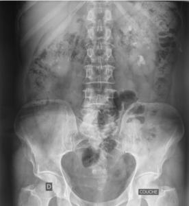
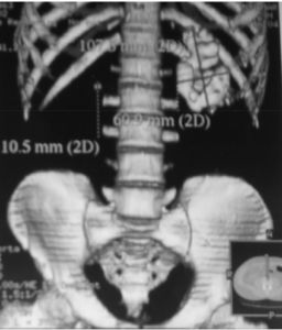
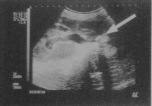
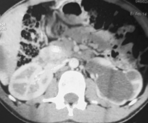
A 30-year-old Woman with a history of diabetes mellitus was followed for 5 years on insulin therapy. The patient presented unilateral lower back pain, severe hydronephrosis and recur- rent urolithiasis. His history was normal until the age of 28, when he experienced episodes of unexplained recurrent fever and chills which were treated with antibiotics. He then re- mained asymptomatic until the age of 29, when he was ad- mitted to the emergency room due to acute unilateral left lower back and iliac pain. The diagnosis revealed appendici- tis treated with antibiotics. Nine months later, she devel- oped new recurrent episodes of urinary tract infection and pyelonephritis accompanied by decreased body weight, se- vere anemia and lower back pain. The renal ultrasound and uroscanner showed severe hydroureteronephrosis with en- larged kidney and multiple urolithiasis (Fig. 1), with no func- tional kidney. The uroscan confirmed a mute kidney on the left and good compensatory function on the right (Fig. 2). The pre- operative diagnosis showed an obstructive megaureter, multi- ple urolithiasis and mute renal (Fig. 3). The abdominal CT scan (Fig. 4) show complete lamination of the renal corte witth sig- nificant distention of the pyelocalicial cavities and purulent contents of the excretory cavities in favor of a destroyed non- secreting sclerotic kidney. The patient felt too anxious to delay treatment for the renal painful.
The clinical diagnosis shows a conscious patient, with fever at 37.8◦C. Biochemical analyzes reveal a creatinine of
14.8 mg/L, normal serum potassium, and a high number of leukocytes. Cytobacteriological examination of urine revealed infection with Candida albicans.
The diagnosis of transit disorders was made by an abdom- inal x-ray which was normal, then an abdominal ultrasound revealed a left pyelocalic dilatation with echogenic content laminating the renal cortex, could be linked to purulent re- tention.
Under ultrasound guidance, the patient underwent a uri- nary diversion. Monitoring of the patient’s condition was marked by apyrexia and a decrease in the number of white blood cells which approached normal values. Kidney function was diagnosed by a kidney scan which showed a normally functioning right kidney and a non-functioning left kidney.
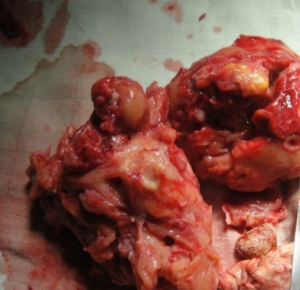
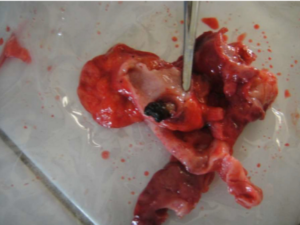
After four weeks, a nephrectomy of the left kidney was performed (Fig. 5). After removing the calculi from the kidney. The stone analysis show monohydrate calcium oxalate (Fig. 6).
Discussion
Urinary stones are solid particles found in the urinary tract, some skiney stones can have a huge size like the bladder stone [4]. They can cause pain, hematuria, nausea, vomiting, and chills with fever related to bacterial urinary tract infections and other symptoms due to several diseases affectinf the local population as neuropsychiatric disorders [5]. Diagnosis is by x-ray imaging, usually a no-exam helical CT scan. Treat- ment is based on analgesics, antibiotics in case of infection, expulsive medical treatment and, sometimes, minimally in- vasive surgical interventions (shock wave lithotripsy or endoscopic stone removal), or by traditional therapy using medic- inal plants [6–8]. Persistence of the stone in the urinary tract can lead to acute renal failure.
Risk factors differ from population to an other. Hypercal- ciuria constituts the main risk factor, it represnts a hereditary condition a hereditary condition which is mainly found in men (75%) compared to women (50%) suffering from cal- cium urinary stones; in the case of a family history of stones, the risk of recurrent stones is increased.
Pyonephrosis is an infection of the upper urinary tract characterized by parenchymal damage and causes subse- quent loss of renal function [9]. It affects all ages and espe- cially young adults [10]. The average age is between 42 and 51 years [11]. It affects both sexes with a female predominance [12]. In 70% of cases, ureteral obstruction and urinary stone are the main anatomical factor in pyonephrosis. Infection is most commonly caused by E. coli, Klebsiella Pseudomonas, Pro- teus, or Enterococcus [13].
Treatment of pyonephrosis includes percutaneous drainage of pus, placement of a retrograde ureteral stent and in extreme cases, nephrectomy. The antibiotics have no effect in pyonephrosis but a surgery intervention is generally necessary. Percutaneous nephrostomy is the most suitable and least invasive method for draining cells retained in the upper urinary tract.
In our case, the diabetic woman was in serious condition with a non-functional kidney, hence the nephrectomy. The diabetes was a risk factor affecting the physiology of organs [14]. In diabetic patients, the clinical presentation and the in- fectious organism may be unusual. Radiological imaging and
uroscanner are means of early diagnosis of this pathology.
Pyonephrosis can be complicated to serious septic shock and sometimes fatal complications. Percutaneous nephrostomy is strongly recommended in the treatment of pyonephrosis and renal scintigraphy is necessary to monitor the functioning of the kidney [15].
Conclusion
Urolithiasis and diabetes coexistence in a patient may com- plicate a pyelonephritis leading to a gigantic pyonephrosis. In this case, which leads to dysfunction of the affected kidney, nephrectomy remains the unique treatment.
Authors’ contributions
MC and AM Collected clinical details, analyzed the data and drafted the manuscript; MC and RAZ were involved in managing the patient; All authors read and approved the final paper.
Patient consent
Written informed consent was obtained from the patient for publication of this case report and any accompanying images. A copy of the written consent is available for review by the Editor-in-Chief of this journal.
- Avnet NL, Roberts TW, Goldberg HR. Tumefactive xanthogranulomatous pyelonephritis. Am J Roentgenol Radium Ther Nucl Med 1963;90:89–96.
- Quinn F, Dick A, Corbally M, McDermott M, Guiney E. Xanthogranulomatous pyelonephritis in childhood. Arch Dis Child 1999;81:483–6.
- Levy M, Baumal R, Eddy AA. Xanthogranulomatous pyelonephritis in children. Etiology, pathogenesis, clinical
and radiologic features, and management. Clin Pediatr (Phila) 1994;33:360–6. doi:10.1177/000992289403300609.
- Chakit M, Aqira A, Mesfioui A. A case report of a giant bladder stone (12 × 8 cm, 610 g). Radiol Case Rep 2024;19(3):970–3. doi:10.1016/j.radcr.2023.11.081.
- Fitah I, Chakit M, El Kadiri M, Brikat S, El Hessni A,
Mesfioui A. The evaluation of the social functioning of schizophrenia patients followed up in the health center My El Hassan of Kenitra, Morocco. Egypt J Neurol Psychiatry Neurosurg 2023;59(1):125. doi:10.1186/s41983-023-00714-7.
- Chakit M, Aqira A, El Hessni A, Mesfioui A. Place of extracorporeal shockwave lithotripsy in the treatment of urolithiasis in the region of Gharb Chrarda Bni Hssen (Morocco). Urolithiasis 2023;51(1):33.
doi:10.1007/s00240-023-01407-9.
- Chakit M, El Hessni A, Mesfioui A. Ethnobotanical study of plants used for the treatment of urolithiasis in Morocco. Pharmacog J 2022;14(5):542–7. doi:10.5530/pj.2022.
- Chakit M, Boussekkour R, El Hessni A, Bahbiti Y, Nakache R, El Mustaphi H, et al. Antiurolithiatic activity of aqueous extract of Ziziphus lotus on ethylene glycol-induced lithiasis in rats. Pharmacog J 2022;14(5):596–602. doi:10.5530/pj.2022.14.141.
- Rabii R, Joual A, Rais H, Fekak H, Moufid K, Bennani S,
et al. Pyonephrosis: diagnosis and treatment: report of 14 cases. Ann Urol (Paris) 2000;34(3):161–4.
- Harrison GS. The management of pyonephrosis. Ann R Coll Surg Engl 1983;65(2):126–7.
- Watt I, Roylance J. Pyonephrosis. Clin Radiol 1976;27(4):513–19. doi:10.1016/s0009-9260(76)80118-3.
- Watson RA, Esposito M, Richter F, Irwin RJ, Lang EK. Percutaneous nephrostomy as adjunct management in advanced upper urinary tract infection. Urology 1999;54(2):234–9. doi:10.1016/s0090-4295(99)00091-6.
- Klein RD, Hultgren SJ. Urinary tract infections: microbial pathogenesis, host-pathogen interactions and new treatment strategies. Nat Rev Micro 2020;18(4):211–26. doi:10.1038/s41579-020-0324-0.
- Baataoui S, Chakit M, Boudhan M, Ouhssine M. Assessment of vitamin D, calcium, cholesterol, and phosphorus status in obese and overweight patients in Kenitra city (Morocco). Res J Pharm Technol 2023;16(7):3405–9.

